|
About
the software
Software Description
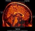
The Brain Atlas Screen Saver/Slideshow presents 10 representative sectionsof
the human brain in coronal sections and 1 sagittal section guide. The "hotmetal"
MRI images are taken every 5 mm anterior to posterior toreveal the most
common brain regions. Presentation rate (in seconds)of each image
is controlled by the user via the Windows® screensaverproperties dialog
box. Labeled regions include: Frontal lobe, Occipital Lobe, Superior
Parietal Gyrus, Calcerine Fissure, Posterior Horn of LateralVentricle,
Cerebellar Cortex, Cerebellar White Matter, Lingula, Calcar Avis, Postcentral
Gyrus, Postcentral fissure, lateral ventricle, cerebellarpeduncle, dentate
nucleus, inferior olive, medulla, flocculus, fourth ventricle,Temporal
lobe, inferior colliculus, splenium of corpus callosum, Parietallobe, precentral
gyrus, inferior parietal gyrus, Superior Temporal Gyrus,Medial Temporal
Gyrus, Inferior Temporal Gyrus, Fusiform Gyrus, Hippocampal Gyrus, red
nucleus, hapenulopeduncluar tract, pulvinar, Cingulate Gyrus, choriod plexus,
pineal, aqueduct, fornix, thalamus, subthalamicnucleus, cerebral peduncle,
hippocampus, pons, interpeduncular fossa, substantia nigra, internal capsule,
habenula, mammillothalamic tract, mammillary body,mediodorsal thalamus,
Insula, Middle Frontal Gyrus, anterior thalamus,anterior hypothalamus,
subiculum, corpus callosum, caudate nucleus, putamen,globus pallidus, claustrum,
extreme capsule, amygdala, septum, externalcapsule, diagonal band, olfactory
sulcus, inferior frontal fissure, genu of corpus callosum, Orbitofrontal
Cortex.
System Requirements
Windows® 9x or higher; 133 MHx Pentium or better;
recommended:SVGA
800 X 600 or higher resolution in High Color mode. |