
|
|
|
|
|
| "The Brain": 9920 |
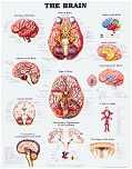
Larger Image
 |
|
| "Anatomy of the Brain": 9921 |
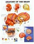
Larger Image
|
The Anatomy of the Brain chart reveals extensive
details of gross structural brain anatomy in awesome perspective views
with rich color. Topics Include: Arteries of the Brain (Base View),
Arteries of the Brain and Neck (Right View), Venous Sinuses, Cerebral Hemispheres
with lateral and sagittal views of structures, Cranial Nerves, Lobes of
the Brain, Meninges and Venous Sinuses, Circulation of the CSF, CSF Pathways
with planes of section, Typical Nerve Cell, Typical Glial Cells. This
chart has some of the best illustrations of the hippocampus and limbic
system available! It also measures 20" wide by 26" high.
Fantastic three dimensional perspectives rendered by Keith Kasnot, C.M.I,
in consultation with Mazen H Khayata, M.D.
|
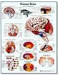
| "Human Brain": VR1615 |
|
|
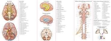
| Anatomy of the Brain Study Guide |
 In Stock Back to Top |
Study Guide Web Price: $9.95 This folding study guide takes our most popular
anatomical images and puts them in a durable, portable format-perfect for
the on-the-go student. Guide shows numbered anatomical structures and contains
answers that can be concealed for easy self-testing and memorization. Write-on,
wipe-off laminated surface. This quick reference covers:
Nerves, arteries, vessels, ventricles and cells, Limbic system and coronal
section, Midbrain, medulla oblongata and spinal cord, Meninges and venous
sinuses, Lobes of the brain, circle of Willis, Somatotopic organization
of the cerebrum
 . . |
| "The Nervous System": 8949 |

Larger Image
|
The Nervous System chart reveals extensive
details of central and peripheral components of the nervous system in rich
color and detail. Topics Include: Brain: Inferior View (cranial
nerves and structures), Peripheral nerves (over 100), Intercostal Nerves,
Midbrain, Medulla Oblongata and Spinal Cord (Posterior View), Spinal Meninges,
Sagittal Section of Female Pelvis. Absolutely
loaded with information. As with our other charts,
this chart measures 20" wide by 26" high. Illustrated by Peter
Bachin. As compared to VR1620, below, this chart shows more detail with
regard to visceral innervation, organization of spinal cord roots, and
pelvic organization of nerves.
|
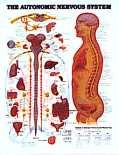
| "The Autonomic Nervous System": 8026 |
Larger Image
|
The Autonomic Nervous System chart reveals
extensive details of central and peripheral components of the Autonomic
nervous system in rich color and detail. Topics Include: Spinal organization
of pre- and postganglionic systems. Target organ innervation from
parasympathetic and sympathetic systems. Organization of preganglionic
parasympathetic
systems in the brainstem -- nuclear groups and cranial nerve components.
Pharmacology (neurotransmitters) of parasympathetic and sympathetic systems.
|
| Anatomy of the Brain Study Guide |
Back to Top |
Web Price: $9.95 This folding study guide takes our most popular
anatomical images and puts them in a durable, portable format-perfect for
the on-the-go student. Guide shows numbered anatomical structures and contains
answers that can be concealed for easy self-testing and memorization. Write-on,
wipe-off laminated surface. This quick reference covers:
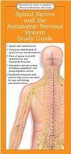
Spinal and cranial nerves, Listing and description of important branches emerging from proximal part of spinal nerves, Spinal cord segments, Descriptions of nerve plexuses, Cutaneous distribution of spinal nerves and dermatomes, View of spinal cord with spinal nerves & immediate branches, Autonomic nervous system, including sympathetic and parasympathetic nerves, Listing of effector organs with sympathetic and parasympathetic action, Folded size approximately 9" x 4". Unfolded size approximately 9" x 24". |
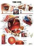
| "The Eye": 9691 |
Larger Image
|
The Eye chart, illustrated by Keith Kasnot,
C.M.I. and Randall Paul, O.D., beautifully illustrates all components of
the eye and connections to the central nervous system. Contents:
External features of the lacrimal glands and nasolacrimal ducts, Central
connections of the visual system, Visual field projection patterns in the
optic nerve and CNS (retinotopic organization), muscles of the eye (abduction
/ accommodation), organization and anatomy of the eye, the Anterior Chamber
systems, The retina, The Fundus, The Macula Lutea, The Lens. As with
our other charts, this chart measures 20" wide by 26" high. Manufactured
by Anatomical Chart Company.
This chart is breathtaking!
-- We keep finding new information every time we pack one for shipment...
|
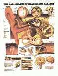
| "The Ear": 9890 |
Larger Image
|
The Ear - Organs of Hearing and Balance chart
is loaded with information pertaining to the external, middle and inner
ear, in addition to the vestibular system. Topics: Tympanic
membrane, Middle ear, Auditory ossicles, Organ of Corti, Membranous Labyrinth,
Membranous Ampulla, Macula of Saccule, How We Hear -- The Physiology of
Sound (EXCELLENT diagrams of Place
Coding at the basilar membrane),
Central Auditory Pathways. As with our other charts, this chart measures
20" wide by 26" high. Another beautiful chart
illustrated by Peter Bachin.
|
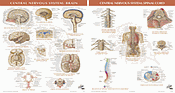
| Netter Charts - Central Nervous System |
|
|
| "Understanding" Clinical Series |

Understanding Alzheimer's Disease 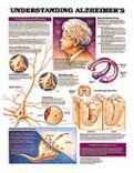
Understanding Parkinson's Disease 
|
|
| The World's Best Anatomical Charts |

Back to Top
|
|
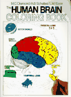
| The Human Brain Coloring Book |
|
|
| Netter's Study Guides and Textbooks |

Our Price: $34.95 |
|

Our Price: $34.95
|
|
| Netter's Atlas of Human Neuroscience |

|
|
Brain Models | Neuroanatomy Charts | Books | Brain Novelty Items | Privacy Policy | Shopping Guarantee | Contact Us...