
|
|
|
|
|
| Skull and Brain Model W-SB |

Decal Info

|
|
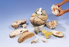
| Human Brain Model CH-1 |

Larger Image Decal Info
|
$29.95 Model Only The human brain Model CH-1 is an inexpensive
brain model that complements general neuroanatomical studies. The human
brain model CH-1 is made of solid acrylic (one of our heaviest models),
and it comes with a sturdy plastic base for storage and display.
Overall, a great brain model for those who need hands-on study of regional
brain anatomy. Painted regions include the thalamus, cerebellum,
and cranial nerves. Additional painting and can be done with colors
of your choice. The three parts shown on the left photo friction
fit with sturdy brass pins to assemble the whole brain shown at right.
No other assembly required. The model can be
labeled using our decal kit. This model does not include a data/diagram
sheet, but brain structures can be easily identified using an introductory
textbook. This model is recommended for self-study only -- For teaching
or presentations we suggest the C-20-c model below. Model;
Brain
Size 5.5" x 4.5" x 4.5".
|
| Brain Model CH-1 Bonus Pack |
|
$46.95 Pack Only Call us crazy...The Bonus Pack
is a bundled package that contains both the Principles
of Psychobiology software and the Human Brain Model Kit CH-1. By
purchasing the Bonus Pack you save 15% on individual products. When
you use the software's programmed system of instruction and the brain model
you'll be VERY proficient in functional neuroanatomy and systems neuroscience.
You can use the software's brain atlas, in combination with its brain images,
to gain 3-dimensional representations of brain structure. The software
also has an electronic "coloring book" so that you can prepare your own
brain coloring book. Hey; it doesn't matter how good the neuroanatomy
imaging software gets...students
always benefit from hands-on experience
with brain models during their studies.
Model; Printed
Info; Diagrams; Brain Size
5.5" x 4.5" x 4.5"Software
(CD-ROM); Single-User License. System Requirements; Windows® 3x; 9x,
ME.
|
| Human Brain Model C20-c |

Larger Image Decal Info
|
|
Larger Images
|
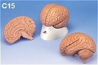
Web Price:$100.00
The human brain model C-15 is a economy life-sized
human brain model with a medial section. Dissectible into 2 parts.
Rigid plastic base included. Lecture-Grade
Model; Printed Info; 6" x 5" x 5"; Student or Patient Use. Compare
the models...
|
Larger Images
|
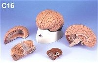
Web Price: $155.00
|
Larger Images
|
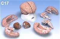
Web Price: $185.00
|

Larger Images
|
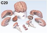
Web Price: $158.00 Cast from a human brain! The human brain model C-18 is a beautiful detailed brain model cast from an actual human brain. The model has very realistic neocortical convolutions and internal structures such as the ventricles. The model rests on a rigid plastic base. This model is especially useful in demonstrating the Hippocampus, lateral ventricles, fornix/fimbria, choriod plexus, etc. Highly recommended for neuroanatomical presentations, clinical or patient use. Visit the Product Page to see Larger Images and labeled structures. |
Larger Images
|
Web Price: $340.00 The human brain model C-20 from 3B is a medially divided deluxe brain model that shows the brain arteries, as well as the removable Circle of Willis. Both halves can be disassembled into Frontal lobe with Parietal lobes, Temporal lobe with occipital lobe, half of brain stem, and half of cerebellum. The model rests on a rigid plastic base. This model is especially useful in clinical settings to clearly demonstrate sources of vascular abnormalities. Highly recommended for clinical, student, or patient use. Visit the Product Page to see Larger Images and labeled structures. |
| C22 - Neuroanatomical Model |

Larger Image
|
Web Price: $305.00 The human brain model C22 from 3B is a high-quality
neuroanatomical model that provides regional color-coding, in addition
to standard numbering/lettering with key card. This
model is especially useful to demonstrate regions of the neocortex such
as primary motor cortex, primary somatosensory cortex, auditory and speech
areas, primary visual cortex, the cingulate, limbic, and infralimbic cortices,
etc. Visit the Product Page
to see labeled structures and larger images. The model is approximately
life-size, and the casting reveals excellent details of sulci, gyri, ventricles
and the fine vermis of the cerebellum.
|
| VH409 2.5X 14-Part Brain Model |
Larger Image
"The VH409 is the ULTIMATE Neuroanatomy training model. Not only are all of the major areas of the brain labeled, but the model is enlarged 2.5X which allows easy visualization of 3-D architecture..." |
Web Price: $679.00 The human brain model VH409 is a ultra-high-quality
2.5X neuroanatomical model with lettering / key booklet. Labeled
structures are: Longitudinal fissure, Superior frontal
sulcus, Inferior fontal sulcus, Central sulcus, Precentral sulcus, Postcentral
sulcus, Intraparietal sulcus, Parietooccipital sulcus, Occipital sulcus,
Superior frontal gyrus, Medial Frontal gyrus, Precentral gyrus, Postcentral
gyrus, Superior parietal lobule, Inferior parietal lobule, Lateral occipital
gyri, Transverse occipital sulcus, Lateral sulcus, Superior temporal sulcus,
Inferior temporal sulcus, Inferior frontal gyrus, Angular gyrus, Supramarginal
gyrus, Superior temporal gyrus, Middle temporal gyrus, Inferior temporal
gyrus, Orgital sulci, Hippocampal sulcus, Calcarine sulcus, Collateral
sulcus, Orbital gyri, Parahippocampal gyrus, Cuneate Lobe, Medial occipitoeemporal
gyrus, Olfactory tract, Olfactory bulb, Straight gyrus, Optic chiasm, Mamillary
body, Mesencephalon, Cingulate sulcus, Sulcus of corpus callosum, Paracentral
lobule, Precuneus, Cingulate gyrus, Cubcallosal area, Rostrum of corpus
callosum, Genu of corpus callosum, Trunk of corpus callosum, Splenium of
corpus callosum, Pellucid septum, Thalamus, Interthlamic connexus, Fornix,
Pituitary gland, Pineal body, Lamina quadrigemina, Forth ventricle, Pons,
Pyramid of medulla, Cerebellum, Lateral ventricle, Insula, Claustrum, Putamen,
Globus pallidus, Amygdala, Hypothalamus, Internal capsule, External capsule,
Extreme capsule, Choriod plexus, Subthalamic nucleus, N. Mamillary body,
Optic tract, Substantia Nigra, Lateral geniculate body, Habenular nucleus,
Uncus of hippocampus, Hippocampus, Pulvinar, Caudate Nucleus, Superior
colliculus, Inferior colliculus, Medial geniculate body, Brachium of Inferior
colliculus, Cerebral crus, Central lobule, Quadrangular lobule, Superior
semilunar lobule, Inferior semilunar lobule, Superior medullary velum,
Vermis of cerebellum, Middle cerebellar peduncle, Dentate nucleus, Fastigial
nucleus, Globose nucleus, Emboliform nucleus, Red nucleus, Cerebral aqueduct,
Nucleus of inferior colliculus, Oculomoter nerve, Trigeminal nerve, Abducens
nerve, Facial nerve, Intermediate nerve, Vestibulocochlear nerve, Glossopharyngeal
nerve, Spinal nerve, Pyramid of medulla, Olive, Anterior median fissure
of spinal marrow, Anterolateral grove of medulla, Anterior funiculus of
cord, Lateral funiculus of cord, Trichlear nerve, Hypoglossal nerve, Vagus
nerve, Accessory nerve, Posterior median sulcus, Superior cerebellar peduncle,
Medial eminence of rhomboid fossa, Facial colliculus, Superior fovea of
rhomboid fossa, Vestibular area of rhomboid fossa, Medullary stria of fourth
ventricle, Inferior fovea of rhomboid fossa, Trigone of vagus, Hypoglossal
trigone, Obex, Posterior intermediate sulcus, Posterolateral groove of
medulla, Fasciculus gracilis of dorsal column, Cuneate fasciculus of dorsal
column -- WOW...It
doesn't get much better than that.! 132 labeled
structures. 34 page manual, including
brain atlas, in 6 languages.
|
| GPI 295 Normal Painted Half-Brain |

|
Web Price: $79.00 The GPI 295 human half-brain model is a new product
that provides color-coding of brain regions, in addition to a laminated
key card. The model is approximately life-size, constructed
of styrene with excellent details of sulci, gyri, 3rd and 4th ventricles,
cerebellar vermis, and sagittal structures. The brain mounts on a
pin boss with a rectangular desk stand. Labeled key card areas are: [Lateral
View] Wernicke's area, Cerebellum, Primary auditory cortex, occipital
lobe and visual association area, angular gyrus (sensory analysis), Parietal
lobe, Primary somatosensory cortex, Motor cortex, Premotor (supplemental
motor) cortex, Prefrontal lobe, Frontal pole (lobe), Broca's area,
Temporal lobe, [Sagittal View] Mammillary body, Olfactory, Optic chiasm,
Anterior commissure, Olfactory bulb, Corpus callosum, Frontal pole (lobe),
Cingulate gyrus, Prefrontal, Supplemental Motor, Motor, Somatosensory,
Parietal lobe, Midbrain, Occipital lobe, Cerebellum, Medulla oblongata.
The model measures approximately 5" d-v, 6.5" a-p, and 2" m-l. Photo key
card includes the above structures. The model itself does not have labels,
but our universal water-slide decal label sheet
can be used to label the model for presentations.
|
| Diseased Brain Model 290 |


Larger Images
|
Web Price: $109.95 The Diseased Brain Model 290 is a full size segmented
brain and skull model. The Brain features a normal hemisphere (half)
and 3 piece coronally-sectioned pathology hemisphere, as well as Circle
of Willis. The brain, which sits inside a partial skull, features
the following pathologies which are also illustrated on a highly-detailed
full-color laminated two-sided education card: alcoholism, Alzheimer's,
aneurism, depression related tumor, seizure related tumor, migraine, multiple
sclerosis, Parkinson's disease, stroke, and subdural hematoma. 5"
x 6-3/4" x 5"
Made of durable high-impact plastic materials. Education card included with model. The skull rests on any horizontal surface. |
| Lecture-Grade Sagittal Head |

Larger Image
|
Web Price: $102.95 The C12 human half-head model is an economically
priced model that demonstrates sagittal structures in addition to 14 bones,
15 structures in the nasal and mouth regions, and 12 structures/muscles
in the neck. With hand-painted labels. Measurements:
26 x 33 x 5 cm. Model and key card.
|
| Basal Skull and Brain Model - 3B |

larger Image
|
Web Price: $367.00 The human brain model C-25 is a highly detailed
model which includes vascular and nervous structures contained in the base
of the skull, in addition to a extremely detailed 8-part brain. The
model includes neocortical surface arteries, subcortical arteries, venous
sinuses, and the internal cartoid. The basal skull includes: CEREBRAL
ARTERIES; All components of Circle of Willis plus opthalamic a.,cerebellar
a., basilar a. labyrinth a. vertebral a. spinal a.; SINUSOF
DURA MATER; Central and PERIPHERAL (peripheral to ganglia; at least
5-10 mm nerve stalks) components of the 12
CRANIAL NERVES including Gasserian ganglion and facial/intermediate
nerves; BONE STRUCTURES include crista frontalis,lamina
cribrosa, anterior cranial fossa, medial cranial fossa, posterior cranial
fossa, vertebra, and CERVICAL SPINAL SEGMENTS.
Over 140 labeled/numbered structures -- See them all at the C25
Product Page. This model is theultimate
resource for anatomy / neuroanatomical training -- the
model has accurate dimensions, accurate color, and it will replace
a cadaver for many studies of the brain and basal skull...Model;
Printed Info; Diagrams; Brain measures 6" x 6" x 6"; Overall Model
Height 11.5" (Skull to Neck).
|
| Head and Brain Models - 3B |

Larger Image |
C10-1 The human head model C10-1 is
a highly detailed model which includes structures contained in the head,
in addition to a extremely detailed 2-part brain. The model includes
neocortical surface arteries, subcortical arteries, venous sinuses, and
the internal cartoid. The head includes structures of the jaw, mouth,
nasal sinuses, skull and internal calvaria. The left eye is removable
for study of the eye muscles and optic nerve. An outstanding model
that allows visualization of the gustatory, olfactory, and optic systems.
See the C10-1 Product Page for Labeled/Numbered
structures. Model; Printed Info; Brain measures 6"
x 6" x 6"; Overall Model Height 10.5" (Skull to Jaw). Compare
all the models...
C11-1 The human head model C11-1 is
a highly detailed model which includes structures contained in the head,in
addition to a lateral view of the left neocortex (not removable). The model
labels the cerebrum and dura mater of the brain. The headincludes
structures of the jaw, mouth, nasal sinuses, and skull. The frontal, shenoidal,
parietal, temporal, occipital, nasal, lacrimal, cheek, superior maxillary,
jaw, and meatus acusticus bones are labeled. The left eye is removable
to study of the eye muscles and optic nerve. Eye structures are eyelid,
palpebralis, ligamentum palpebrale mediale, lacrymalsac, m. levator palpebrae
sup., tendon of m. obliquus sup., sup. rectusmuscle, lateral rectus, muscle,
inferior rectus muscle, medial rectus muscle, m. obliquus inferior muscles
and optic nerve. 26 labeled/numbered regions
in total. Model;
Printed Info; Overall Model Height 10.5". Compare
all the models...
Sheep Brain Dissection Packages |
Brain Models | Neuroanatomy Charts | Books| Brain Novelty Items | Privacy Policy | Shopping Guarantee | Contact Us...
Copyright©1998-2006, Brain-Mart, An Internet Division of Red Reef
Publications
P.O. Box 4286, Boynton Beach, FL 33424-4286, FAX 561-735-3856
